Early detection is the single most important factor in improving outcomes for people with oral cancer. When abnormal tissue is identified at an early, localized stage, treatment options are broader and the chances of long-term survival are substantially higher. Routine screening gives clinicians the opportunity to catch subtle changes before they progress, which is especially important because early lesions are often painless and easy to overlook.
Oral cancer can affect the lips, tongue, floor of the mouth, and other soft tissues inside the mouth. While it is less common than some other cancers, its impact is significant because late-stage disease frequently requires more extensive treatment and leads to a heavier toll on function and quality of life. That’s why integrating dependable screening tools into regular dental visits is a practical, proactive step toward better health.
Screening is not about causing alarm — it’s about evidence-based vigilance. A consistent, systematic approach to oral examinations helps clinicians establish a baseline for what is normal in each patient. From that baseline, any new, persistent, or suspicious change can be recognized and acted on quickly, with appropriate follow-up and specialty referral when indicated.
VELscope® is a handheld diagnostic aid that uses a specific wavelength of blue light to illuminate the oral mucosa. Healthy tissue fluoresces in predictable ways, while areas that have undergone cellular or structural changes often show altered fluorescence patterns. By highlighting these differences, VELscope® helps clinicians visualize abnormalities that may be indistinguishable under regular lighting.
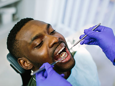
The examination is straightforward and noninvasive. In a dimmed operatory, the clinician directs the light to different areas inside the mouth and observes the fluorescence response. Areas that look atypical are noted for closer clinical evaluation. Importantly, VELscope® is an adjunctive tool — it does not replace a thorough visual and tactile exam, nor does it by itself provide a definitive diagnosis.
Because the device is portable and quick to use, it can be integrated into routine hygiene and wellness visits without significant disruption. Its real value lies in its ability to direct attention to subtle changes early, supporting the clinician’s judgment about when to monitor, document, or refer for additional testing.
A typical VELscope® screening adds only a few minutes to a regular dental checkup. After a standard oral exam and discussion of any symptoms or risk factors, your provider will dim the lights and use the device to scan the oral tissues. There’s no contact with sensitive equipment and no discomfort — patients simply look into the light while the clinician methodically evaluates each area.
Findings are documented in the chart, and the clinician explains what they observed in plain language. If an area appears suspicious under fluorescence, the team will compare it with the visible appearance and consider factors like duration, texture change, and risk profile. Many suspicious findings merit a conservative watch-and-wait approach with short-term rechecks; others may require referral to an oral surgeon or ENT for biopsy.
Preparation is minimal — there’s no special diet, medication change, or advance testing required. For patients with risk factors such as tobacco or heavy alcohol use, a history of HPV exposure, or unexplained oral sores, incorporating VELscope® into regular visits offers an added layer of safety and reassurance.
VELscope® brings clear advantages: it increases the clinician’s ability to detect subtle tissue changes and complements a thorough clinical examination. In many cases, fluorescence screening prompts closer observation or earlier referral, which can lead to more timely diagnosis. For practices focused on prevention and early detection, the technology is a useful extension of traditional screening methods.
That said, fluorescence screening has limitations that are important to understand. Not every area that appears different under blue light is cancerous; inflammation, trauma, and benign lesions can also alter fluorescence. Conversely, a normal fluorescence pattern does not guarantee the absence of disease. Because of these factors, VELscope® findings must be interpreted within the context of a full clinical assessment, patient history, and, when necessary, tissue biopsy.
A pragmatic approach recognizes VELscope® as a sensitive but not exclusively specific tool. Used thoughtfully, it reduces the chance of missed lesions and helps prioritize cases that merit further investigation, while avoiding overdiagnosis through careful clinical correlation and follow-up planning.
At Cherokee Smiles Dental, we view oral cancer screening as an essential component of comprehensive dental care. Our clinicians incorporate VELscope® examinations into routine checkups for patients based on individual risk and clinical findings, ensuring that each visit addresses both immediate oral health needs and long-term wellness. This integrative model helps us detect changes early and coordinate timely next steps when needed.
If a suspicious area is identified, our team follows a clear pathway: documentation, patient education, short-interval monitoring when appropriate, or referral to a specialist for diagnostic biopsy. We prioritize clear communication so patients understand why a follow-up is recommended and what the potential outcomes may be, without creating unnecessary worry.
Beyond the technology itself, we emphasize prevention through education. We talk with patients about modifiable risk factors, how to perform basic self-checks at home, and when to report new symptoms. Our goal is to create a partnership around oral health that empowers patients and supports early intervention whenever it’s warranted.
In summary, VELscope® cancer screening is a practical, noninvasive adjunct to the standard oral exam that can help clinicians spot suspicious tissue changes sooner. When combined with a thorough clinical evaluation and sensible follow-up, it strengthens a prevention-focused approach to oral health. If you have questions about how VELscope® screenings are used in our practice or whether this test is right for you, please contact us for more information.


VELscope® cancer screening is an adjunctive diagnostic exam that uses a specific wavelength of blue light to evaluate the oral mucosa for altered tissue fluorescence. The device highlights areas where cellular or structural changes have disrupted normal tissue fluorescence, allowing clinicians to see patterns that can be hard to detect under regular lighting. It is designed to complement, not replace, a thorough visual and tactile oral exam performed by a dental professional.
The screening is quick and noninvasive and is commonly used during routine dental checkups to add an extra layer of surveillance. Findings are documented and interpreted in the context of the full clinical examination and patient history. When warranted, abnormal findings prompt closer monitoring or referral for further diagnostic testing.
VELscope® works by emitting a blue light that causes healthy oral tissues to fluoresce in predictable patterns, while tissues with structural or cellular changes often show altered or diminished fluorescence. These fluorescence differences create visual contrast between normal and suspicious areas, guiding the clinician to regions that may need closer inspection. The technique can reveal subtle changes in the mucosa before they become obvious to the naked eye.
During the exam the operatory lights are dimmed and the clinician scans the mouth systematically, observing fluorescence responses across lips, tongue, floor of mouth, and other soft tissues. Any atypical fluorescence is compared with the visible appearance, patient history, and known risk factors. This targeted visualization helps prioritize areas for monitoring, documentation, or referral for biopsy when clinically indicated.
The VELscope® exam is noninvasive and generally comfortable for most patients, adding only a few minutes to a routine dental visit. The procedure involves no tissue contact with sensitive instruments and causes no pain; patients simply look toward the light while the clinician evaluates different areas of the mouth. There is no need for anesthesia, special preparation, or recovery time after the screening.
Because the exam is quick and portable it can be integrated into hygiene or wellness visits with minimal disruption. Patients who experience strong gag reflexes or other specific sensitivities should mention that to the clinician so accommodations can be made. As with any clinical test, the clinician will explain findings in plain language and outline next steps if something of concern is detected.
VELscope® screening is a valuable adjunct for many patients, particularly those with known risk factors such as tobacco use, heavy alcohol consumption, a history of HPV exposure, a history of previous oral lesions, or persistent oral sores that do not heal. It is also useful as part of routine surveillance for patients with a history of oral premalignant conditions or prior head and neck cancer. For low-risk individuals, the tool can still provide reassurance and help establish a baseline of normal tissue appearance.
How often screening is performed depends on individual risk and clinical judgment; many practices incorporate VELscope® into annual or biannual exams for higher-risk patients and evaluate its use case-by-case for others. Frequency may be increased when a patient has persistent symptoms or when clinicians want short-interval surveillance of a suspicious finding. Your dental team will recommend an appropriate schedule based on your medical history and clinical findings.
An abnormal VELscope® finding means the tissue is exhibiting altered fluorescence compared with surrounding healthy tissue, which can indicate a range of conditions from benign inflammation or trauma to dysplasia or malignancy. Because altered fluorescence is not specific to cancer, clinicians use these findings as a cue to perform a more detailed clinical assessment rather than as a standalone diagnosis. The next step is always correlation with visual and tactile exam results and the patient’s risk profile.
When an area appears atypical, the team documents its appearance, discusses the observation with the patient, and determines whether short-interval rechecks, photographic monitoring, or referral for specialist evaluation and biopsy are appropriate. Conservative watchful waiting with scheduled follow-up is common for lesions that do not have clear worrisome features, while persistent or progressive changes prompt expedited referral. Clear communication ensures patients understand the rationale for any recommended follow-up.
No, VELscope® cannot by itself determine whether tissue is cancerous; it is an adjunctive screening tool that highlights areas of altered fluorescence that warrant further evaluation. A definitive diagnosis of cancer requires histopathologic confirmation via biopsy and laboratory analysis. The value of VELscope® is in helping clinicians detect and prioritize suspicious areas that might otherwise be overlooked during a routine exam.
When the VELscope® exam identifies a suspicious area, clinicians integrate that information with the physical exam, clinical history, and risk factors to decide on the appropriate next steps. If clinical concern remains high after correlation, referral to an oral surgeon, ENT specialist, or oral pathologist for biopsy is the standard course to establish a diagnosis and guide treatment.
Preparation for a VELscope® screening is minimal; there is no need for fasting, medication changes, or special dietary restrictions prior to the exam. Patients should arrive as they would for a routine dental visit and be ready to share relevant medical and social history, including tobacco or alcohol use, prior oral lesions, and any recent changes in symptoms. Providing an accurate history helps the clinician interpret fluorescence findings in the correct clinical context.
If you have had recent dental procedures, oral ulcers, or topical treatments that could affect mucosal appearance, mention those to your provider so they can factor them into their assessment. Bring up any persistent sores, lumps, numbness, or changes in sensation you have noticed, since these symptoms guide the exam and follow-up recommendations. Clear communication makes the screening more informative and efficient.
VELscope® is a sensitive adjunctive tool that can increase detection of subtle mucosal changes when used together with a thorough visual and tactile exam, but it is not a replacement for traditional clinical assessment. The device tends to be sensitive to abnormal tissue changes, which helps reduce the chance of missing lesions, but it is less specific because benign conditions such as inflammation or trauma can also alter fluorescence. For that reason, VELscope® results must be interpreted within the broader context of the patient’s exam and history.
In practical terms, VELscope® enhances clinical vigilance by pointing clinicians toward areas that merit closer inspection, documentation, or monitoring. It can improve early detection when used thoughtfully, but clinicians rely on their comprehensive assessment and, when needed, biopsy confirmation to make definitive diagnostic and treatment decisions. This balanced approach helps avoid unnecessary procedures while improving the chance of timely intervention for concerning lesions.
At Cherokee Smiles Dental, VELscope® screening is offered as part of an integrative approach to oral health and cancer surveillance, incorporated into routine checkups based on individual risk and clinical findings. The tool is used to supplement the standard oral cancer exam, helping our clinicians identify subtle changes earlier and document baselines for ongoing comparison. The screening is performed in our Woodstock, GA office during a regular visit when clinically appropriate.
If a suspicious area is detected, our team follows a clear pathway that includes documenting the finding, educating the patient about its significance, arranging short-interval follow-up when appropriate, and coordinating referrals for specialist evaluation or biopsy as needed. We emphasize patient communication and preventive education so you understand the recommended next steps and what to watch for between visits. This coordinated approach supports timely intervention and continuity of care.
Regular self-observation of the mouth can complement clinical screenings by helping you notice new or changing areas between appointments; look for persistent sores, white or red patches, lumps, unexplained pain, numbness, or difficulty swallowing. Performing a simple monthly self-check in front of a mirror under good lighting and reporting any persistent changes to your dental team can help with early detection. Keep a record or take photos of any concerning areas so you can show changes over time.
Reducing modifiable risk factors also supports oral health and lowers cancer risk; quitting tobacco, limiting heavy alcohol use, maintaining good oral hygiene, and discussing HPV vaccination with your medical provider are practical steps. Share any changes in your general health or habits with your dentist, since those details affect screening frequency and follow-up decisions. Partnering with your dental team and staying proactive between visits strengthens an early-detection strategy.
Need to schedule an appointment or have questions about our services?
Our friendly team makes it easy to get the care you need. Whether you call or submit our online form, we’re here to guide you every step of the way.
Don’t wait to start your journey to a healthier, brighter smile—contact us today and experience personalized dental care you can trust.