An intraoral camera is a compact, pen‑sized imaging device designed to capture clear, full‑color images from inside the mouth. Equipped with a small lens and often LED illumination, the camera produces high‑resolution photos and live video that can be displayed instantly on a chairside monitor. This direct visual access allows dental teams to examine teeth, restorations, and soft tissues with a level of detail that is difficult to achieve with the naked eye alone.
Unlike traditional mirrors or tactile exploration, the intraoral camera magnifies and documents findings, making even subtle surface changes visible. The device is maneuverable and gentle; most patients feel only minimal awareness of the camera’s presence while images are captured. Because these images are digital, they can be enhanced, annotated, and archived with relative ease to support clinical decisions.
From routine preventive visits to more complex restorative planning, intraoral imaging serves as a straightforward way to translate clinical observations into objective, visual records. For patients, this means a clearer understanding of oral health; for clinicians, it provides reliable evidence to guide diagnosis and treatment discussions.
One of the primary advantages of intraoral imaging is improved diagnostic clarity. High‑resolution photos reveal cracks, early decay, margin defects, and soft tissue changes that might be missed during a cursory exam. When a suspected problem is captured on camera, the dental team can evaluate it from multiple angles, document progression over time, and determine whether additional testing or intervention is warranted.
Equally important is the way these images support communication. Patients often struggle to interpret technical descriptions of a condition; seeing a large, color image of the affected area makes explanations immediate and accessible. This shared visual reference reduces uncertainty, encourages questions, and helps patients make informed choices about their care.
For the clinician, intraoral photos also facilitate clearer handoffs to specialists and laboratories. A well‑composed image communicates details about tooth orientation, shade, and restoration margins that written notes alone might not convey. That clarity supports coordinated care and reduces the likelihood of misinterpretation during treatment planning.
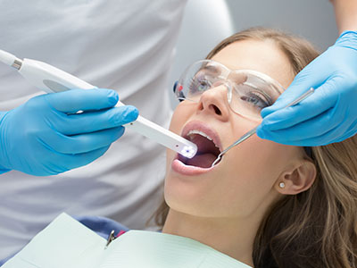
An intraoral camera exam is typically quick, painless, and noninvasive. During a routine checkup or when evaluating a specific concern, your dental provider will position the camera inside the mouth to capture still images or short video clips. The camera’s small size and bright light allow fast, well‑illuminated shots without discomfort, and most images are captured in seconds.
While images are being taken, they are usually displayed on a nearby monitor so patients can follow along in real time. This live viewing experience provides an immediate visual explanation of what the clinician is seeing and why certain areas may need attention. If a patient prefers, the clinician can pause to explain findings or save selected images for later review.
After the exam, saved images become part of the patient’s digital chart. These records can be revisited during future visits to compare changes, demonstrate healing, or show how a restoration is holding up. The process is straightforward, and many patients appreciate the transparency and education that intraoral imaging offers.
Modern intraoral cameras are designed to integrate seamlessly with digital practice management and imaging systems. Captured images can be tagged, stored, and organized within a patient’s electronic record, providing a chronological visual history that complements radiographs and clinical notes. This integration makes it easier to track the progression of lesions, restorations, and periodontal conditions over time.
In treatment planning, intraoral photos serve multiple purposes: they help document baseline conditions, verify the fit and aesthetics of prosthetics, and supply precise visual information to dental laboratories. When crowns, veneers, or other restorations are fabricated, photographs showing tooth shade and contour are invaluable in achieving predictable, aesthetic outcomes.
Digital imaging also streamlines administrative tasks. When specialists, insurance carriers, or labs request documentation, clinicians can provide clear, annotated images that support the clinical rationale for a proposed treatment. This reduces back‑and‑forth and helps ensure that collaborators have the details they need to proceed confidently.
Intraoral cameras are designed with patient safety and infection control in mind. Many models use disposable sheaths or autoclavable tips to prevent cross‑contamination between patients, and clinicians follow standard sterilization protocols to ensure cleanliness. The noninvasive nature of the device means there is no tissue disruption, and images are obtained without ionizing radiation.
Clinically, intraoral imaging supports early intervention. By documenting small defects or early caries that might otherwise go unnoticed, clinicians can recommend conservative treatments that preserve more of the natural tooth structure. In periodontal care, photographs help visualize soft tissue inflammation, recession, and plaque accumulation, enabling targeted hygiene instruction and monitoring.
Beyond diagnostics, intraoral cameras enhance patient engagement and case acceptance by making treatment needs transparent. When patients can see what the clinician sees, they often feel more involved in their care plan and more confident in the recommended next steps. That collaborative approach supports better outcomes and a more positive patient experience.
Intraoral cameras are a practical, patient‑centered technology that brings clarity to clinical exams, strengthens communication, and integrates smoothly with modern digital records. At Cherokee Smiles Dental in Woodstock, we use intraoral imaging as part of a thoughtful diagnostic process to help patients understand their oral health and make informed decisions. If you’d like to learn more about how intraoral cameras fit into your exam or treatment plan, please contact us for more information.


An intraoral camera is a compact, pen-sized imaging device that captures full-color photographs and short video of the teeth and soft tissues inside the mouth. The device uses a small lens and LED illumination to produce high-resolution images that are displayed on a chairside monitor in real time. Because the camera is maneuverable and slender, clinicians can visualize areas that are difficult to see with the naked eye.
The images are digital and can be enhanced, annotated, and stored within the patient record to support diagnosis and treatment planning. Capture is usually noninvasive and completed in seconds, with minimal patient awareness of the device. This direct visual record transforms subjective observations into objective documentation for future comparison.
High-resolution intraoral photos can reveal small cracks, early decay, margin defects, and soft tissue changes that may be missed during a cursory visual exam. Multiple angles and magnification allow the clinician to assess surface texture, restoration margins, and subtle color changes that inform diagnostic decisions. When findings are documented, clinicians can track progression over time and determine whether conservative or more advanced intervention is warranted.
Images also support predictable treatment planning by providing clear visual references for lab communication and specialist referrals. Photographs that show tooth orientation, shade, and surrounding tissues reduce ambiguity when fabricating crowns, veneers, or other restorations. This visual evidence helps clinicians and patients set realistic expectations and verify outcomes after treatment.
An intraoral camera exam is typically quick, noninvasive, and comfortable for most patients. The clinician or assistant will position the camera inside the mouth and capture still images or short video clips while the images are displayed on a nearby monitor. Most images are obtained within seconds and require only minimal cooperation from the patient.
Patients are often invited to view images as they are taken so the clinician can explain findings in real time and answer questions. Selected images are saved to the patient’s electronic chart for future reference and comparison. This immediate visual feedback enhances understanding and supports shared decision making.
Yes, intraoral images are stored digitally within the patient’s electronic record as part of their clinical documentation. Modern practice management and imaging systems allow images to be tagged, annotated, and organized so clinicians can build a chronological visual history. These records make it easy to compare baseline conditions with later visits to document healing, progression, or the performance of restorations.
Stored images can also be used for referrals and lab communication when additional detail is needed to plan treatment. Clinicians follow standard policies for record retention and privacy to protect patient information. Access to images is governed by the practice’s data management protocols and applicable privacy regulations.
An intraoral camera captures surface and soft tissue detail in full color, while dental X-rays reveal internal structures such as tooth roots, bone levels, and interproximal decay. The two technologies are complementary rather than interchangeable; intraoral photos show texture, margins, and color, whereas radiographs provide information about what lies beneath the surface. Clinicians typically use both tools together to form a complete clinical picture.
Unlike X-rays, intraoral cameras do not expose patients to ionizing radiation and are ideal for documenting visible conditions and monitoring soft tissue changes. Radiographs remain necessary for diagnosing issues that cannot be seen externally, such as bone loss or deep decay. Together, they provide a more accurate basis for diagnosis and treatment planning.
Intraoral cameras are designed with infection control in mind and are used with disposable sheaths or autoclavable tips to prevent cross-contamination. Clinicians follow standard sterilization and personal protective equipment protocols to maintain a safe clinical environment. The imaging process itself is noninvasive and does not disrupt tissues.
Because the device does not use ionizing radiation, it is safe for repeated use during routine exams and follow-up visits. Proper handling and adherence to manufacturer cleaning guidelines preserve device performance and patient safety. If you have specific concerns about infection control, your dental team can explain the steps they take to protect patients.
Yes, intraoral photographs are a valuable tool for communicating with specialists and dental laboratories because they convey precise visual details that written notes alone may not capture. Images can show tooth shade, contour, margin location, and gingival architecture, which helps laboratories create restorations that match the clinical situation. When referring a patient, clinicians can include annotated photos to clarify findings and streamline coordination of care.
Providing clear, well-composed images reduces the likelihood of misinterpretation and accelerates decision making between providers. Photographic documentation also supports treatment authorization and clarifies the clinical rationale when additional input is requested. Secure transmission and appropriate consent are used when sharing images outside the practice.
While intraoral cameras excel at documenting surface detail and soft tissue appearance, they cannot visualize internal tooth structure, bone density, or conditions beneath restorations like radiographs can. Lighting, moisture, and limited access in certain areas of the mouth can sometimes affect image quality. Capturing consistently diagnostic photographs requires training and proper positioning techniques.
Images are a supplement to, rather than a replacement for, other diagnostic tools such as clinical probing and radiographic imaging. Clinicians interpret photographs in the context of a full clinical exam and diagnostic tests to avoid over-reliance on a single information source. When limitations exist, additional imaging or specialist consultation may be recommended.
Seeing a clear, enlarged image of a specific tooth or area makes clinical observations more tangible and easier to understand than verbal descriptions alone. Visual evidence reduces uncertainty, prompts focused questions, and helps patients grasp why certain treatments are advised. This transparent approach fosters informed consent and more active participation in care decisions.
Saved images provide a reference that patients and clinicians can revisit to monitor healing or the progression of a condition over time. Comparing images from different visits helps illustrate the effects of treatment or changes in oral hygiene practices. The visual record also supports patient education by highlighting areas where home care can make a measurable difference.
At Cherokee Smiles Dental in Woodstock, intraoral cameras are used during exams to document findings, educate patients, and support treatment planning. The device enables clinicians to show patients what they see, archive baseline images, and communicate exact details to laboratories or specialists when necessary. Images are incorporated into the electronic chart so they are available for follow-up comparison and collaborative care.
This technology is applied across preventive, restorative, and periodontal visits to enhance diagnostic clarity and patient engagement. Clinicians combine intraoral imaging with other diagnostic tools such as radiographs and clinical probing to develop a comprehensive treatment approach. If you have questions about how images will be used in your care, the team can explain the process and answer any privacy or technical concerns.

Need to schedule an appointment or have questions about our services?
Our friendly team makes it easy to get the care you need. Whether you call or submit our online form, we’re here to guide you every step of the way.
Don’t wait to start your journey to a healthier, brighter smile—contact us today and experience personalized dental care you can trust.