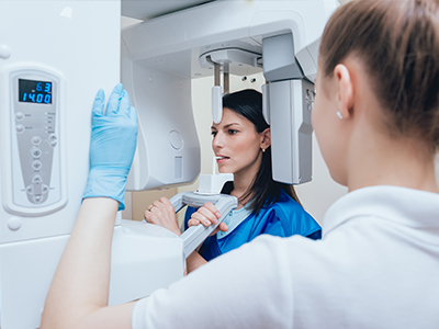
Digital radiography replaces traditional film with electronic sensors and computer processing to capture dental X-rays. Instead of waiting for film to develop, images appear almost instantly on a monitor, allowing clinicians to review and interpret findings in real time. This shift from chemical-based imaging to digital workflows has reshaped how dentists evaluate tooth structure, bone levels, and restorations.
Beyond speed, digital radiography improves the practical side of diagnosis. Images can be enhanced, magnified, and adjusted for contrast and brightness without retaking exposures, which helps clinicians spot subtle changes earlier. That can translate into more precise treatment planning and clearer communication between the dental team and patients during office visits.
For patients, the most noticeable differences are improved comfort and a more efficient visit. Because images are available immediately, consultations and next steps happen faster, and the practice can spend more time discussing findings and treatment options rather than waiting on film development.
Digital systems rely on a variety of sensor technologies to produce diagnostic-quality images. Direct-capture sensors (often CMOS or CCD based) convert X-rays into electronic signals instantly, while phosphor plate systems store an image that is later scanned into the computer. Each approach has benefits: direct sensors deliver immediate images with high resolution, and plate systems offer a flexible, thin option that many patients find comfortable.
Once an image is captured, software tools enhance its diagnostic value. Clinicians can zoom in to inspect tiny details, adjust grayscale to reveal hidden contrasts, and apply measurement tools for evaluating bone levels or root lengths. These capabilities reduce guesswork and help clinicians make more informed decisions about restorative or periodontal care.
Sensor design has also improved patient ergonomics. Newer sensors are smaller and more contoured, minimizing gag reflexes and discomfort during bitewing and periapical imaging. Faster capture times and reduced need for repeats make the experience quicker and less stressful for children and adults alike.
One of the most important advantages of digital radiography is reduced radiation exposure compared with traditional film X-rays. Digital detectors are more efficient at capturing X-ray photons, which allows clinicians to use lower settings while maintaining image quality. Dentists follow the ALARA principle—keeping doses As Low As Reasonably Achievable—by tailoring exposures to the diagnostic need for each patient.
Safety measures extend beyond equipment selection. Proper shielding, careful sensor placement, and using the smallest field-of-view necessary for a specific diagnostic task all contribute to minimizing exposure. Pediatric patients and individuals with special health considerations receive extra attention so imaging is both safe and effective.
If you have specific concerns about radiation, your dental team can explain why an X-ray is recommended and describe the precautions taken. In many cases, the diagnostic benefit of a targeted radiograph outweighs the minimal risk, because the information gained guides timely and appropriate care.
Digital images integrate directly with electronic dental records, which streamlines clinical workflows and reduces paperwork. Instead of filing physical films, teams store images within the patient chart where they are indexed, easy to retrieve, and available during every visit. This efficiency helps ensure nothing is overlooked when reviewing a patient’s history or planning multi-stage treatments.
The ability to share high-resolution images instantly enhances collaboration with specialists and labs. Whether coordinating a referral for oral surgery, sending images for implant planning, or communicating with a dental laboratory about a crown, digital files can be transmitted securely and reviewed by multiple providers simultaneously. That coordination supports more accurate, predictable outcomes.
Digital radiographs also improve patient education. By displaying images on a chairside monitor, clinicians can point out areas of concern and walk patients through the visible anatomy. Seeing an enlarged image helps many patients understand recommended care and makes informed decisions easier during the consultation.
At Cherokee Smiles Dental, this connected workflow supports a smoother patient experience: from diagnosis to treatment planning and follow-up, digital imaging helps our team work efficiently on behalf of each patient.
Storing and managing digital radiographs responsibly is a critical part of modern dental practice. Secure servers, encrypted backups, and strict access controls help protect patient records from loss or unauthorized access. Practices that handle images professionally also follow applicable privacy rules and industry best practices to safeguard sensitive health information.
Integration with other digital tools amplifies the value of radiography. When radiographs work alongside intraoral cameras, digital impressions, and diagnostic software, clinicians gain a fuller picture of oral health. This integrated approach supports more precise restorations, better monitoring of periodontal conditions, and improved long-term maintenance plans.
Reducing repeat exposures is another practical benefit: because digital images are immediately reviewable and easily enhanced, fewer retakes are required. That reduces cumulative exposure for patients and saves time for the dental team, allowing visits to focus on treatment and education instead of repeating procedures.
Whether used for routine exams, restorative planning, or monitoring healing after treatment, digital radiography is an essential tool in contemporary dentistry. It supports clearer diagnoses, safer imaging practices, and a more connected standard of care across providers.
In summary, digital radiography brings faster results, improved image quality, and enhanced safety to routine dental imaging. The technology supports clearer communication among clinicians and with patients while integrating seamlessly into modern treatment workflows. If you’d like to learn more about how digital imaging is used during visits to Cherokee Smiles Dental or how it may benefit your care, please contact us for more information.


Digital radiography uses electronic sensors and computer processing to capture dental X-rays instead of traditional film, producing images almost instantly for review. This technology lets clinicians view, enlarge, and enhance images in real time, improving the ability to detect decay, bone changes, and issues around restorations. Faster image availability supports more efficient consultations so patients spend less time waiting and more time discussing care options.
Because images can be stored and retrieved electronically, digital radiography also streamlines record keeping and follow-up. Enhanced images reduce the need for repeat exposures and support clearer communication between the dental team and patients. Many practices, including the office of Cherokee Smiles Dental, integrate these images directly with patient charts to improve continuity of care.
Unlike film X-rays that require chemical processing, digital radiography captures images electronically and displays them on a monitor within seconds. Digital systems allow clinicians to adjust contrast, zoom, and apply measurement tools without retaking images, which improves diagnostic accuracy. The immediate feedback reduces workflow delays and often shortens appointment time for patients.
Digital files are also easier to archive and transmit compared with physical films, removing the need for manual storage and retrieval. Because detectors are more efficient, exposures can typically be lower while maintaining diagnostic quality. These differences make digital radiography more flexible and better suited to modern, integrated dental workflows.
Digital radiography generally exposes patients to lower radiation doses than traditional film because modern detectors capture X-rays more efficiently. Dentists follow the ALARA principle — keeping radiation As Low As Reasonably Achievable — by selecting the smallest field of view and the lowest exposure settings necessary for diagnosis. Proper shielding and sensor placement further reduce any radiation that reaches the patient.
Pediatric patients and individuals with special health considerations receive tailored protocols to minimize exposure while still obtaining essential diagnostic information. If you have specific concerns about radiation, your dental team can explain why an X-ray is recommended and describe the safety steps used during your visit. In most cases, the diagnostic benefit of targeted imaging outweighs the minimal risk, helping to prevent or treat oral health problems earlier.
Digital systems typically use direct-capture sensors such as CMOS or CCD devices and indirect-capture phosphor plate systems that are scanned into the computer. Direct sensors deliver immediate, high-resolution images and are often used when rapid feedback is needed, while phosphor plates are thin, flexible, and sometimes more comfortable for patients. Extraoral sensors, such as panoramic or cone beam units, are used when broader views of the jaws and facial structures are required.
Each sensor type has clinical advantages, and clinicians choose the best option based on diagnostic need, patient comfort, and the area being imaged. Advances in sensor design have reduced bulk and improved ergonomics, which helps minimize gag reflex and discomfort during intraoral exposures. The result is clearer images with fewer repeats and a more comfortable experience for patients of all ages.
Digital radiographs reveal details that are not visible during a visual exam, such as early interproximal decay, root infections, bone loss from periodontal disease, and the status of previous restorations. Software tools allow clinicians to enhance contrast, measure distances, and zoom in on areas of concern, which supports more precise diagnosis and treatment planning. These capabilities are particularly useful for endodontic assessments, implant planning, and detecting hidden fractures or pathology.
Because images can be compared over time, digital radiography also supports monitoring of healing and disease progression with greater consistency. Clinicians can store baseline images and reference them at follow-up visits to evaluate changes, adjust care plans, and document treatment outcomes. This longitudinal view helps ensure timely interventions and better long-term oral health management.
A typical digital X-ray appointment begins with the clinician explaining the reason for the image and positioning a small sensor or plate in the mouth for intraoral views, or guiding the patient for an extraoral exposure such as a panoramic shot. Exposure times are very short, and modern sensors capture images swiftly so the process is quick and comfortable. Protective measures like a lead apron and thyroid collar may be used based on clinical judgment and patient preferences.
After the exposure, the image appears on a monitor almost immediately and the clinician can review it with the patient, pointing out findings and next steps. If additional views are needed, software adjustments often resolve issues without repeating exposures. The overall experience is designed to be efficient and minimally invasive for patients.
Responsible image management includes secure storage on encrypted servers, regular backups, and restricted access controls to protect patient privacy and maintain data integrity. Many modern dental offices integrate radiographs with electronic health records and follow industry best practices and applicable privacy regulations to safeguard sensitive information. Access is typically limited to authorized clinicians and staff who need the images for diagnosis and treatment.
Secure transmission protocols also enable safe sharing when coordination with specialists or labs is necessary, while audit trails document who accessed an image and when. Patients can also request copies of their images for continuity of care, and the practice will provide them through secure channels to preserve confidentiality. These measures help ensure that digital radiographs remain both accessible for care and protected against unauthorized use.
Yes, digital radiographs are easily shared with specialists, oral surgeons, and dental laboratories using secure electronic transfer methods that preserve image quality. Sharing images expedites referrals, collaborative treatment planning, and laboratory communication for restorations, which supports more predictable outcomes. Clinicians can send high-resolution files along with clinical notes so consulting providers have the context they need to make recommendations.
The ability to transmit images quickly reduces delays associated with mailing physical films and allows multiple providers to review the same files simultaneously. When coordinating complex care, this streamlined exchange improves accuracy and helps ensure that treatment steps align across the care team. Patients who are referred for specialist care often benefit from faster, better-informed consultations because images travel with the clinical record.
Children and pregnant patients are managed with additional caution and individualized protocols to minimize exposure while still obtaining necessary diagnostic information. For pediatric patients, clinicians adjust exposure settings, limit the number of views to those that are essential, and use the smallest field of view possible to answer the clinical question. For pregnant patients, X-rays are only performed when absolutely necessary and after discussing alternatives and timing with the patient and care team.
Protective measures such as lead aprons and thyroid collars are commonly used for these groups, and clinicians carefully document the diagnostic need for any exposure. If an X-ray can be safely deferred or an alternative diagnostic approach used, the dental team will discuss those options with the patient. The goal is to balance diagnostic benefit with prudent safety considerations for every individual.
The frequency of dental X-rays depends on each patient's oral health, risk factors, and the clinical questions the dentist needs to answer. New patients often receive baseline images to establish a diagnostic reference, while patients with active disease, ongoing treatment, or higher risk for decay may need more frequent monitoring. Conversely, low-risk patients with stable oral health may require fewer images spaced farther apart.
Dental teams evaluate factors such as age, medical history, past radiographic findings, and current symptoms when recommending imaging intervals. Decisions about timing and the specific type of radiograph are individualized to ensure images are obtained only when they will meaningfully affect diagnosis or treatment. If you have questions about why an X-ray is recommended, your dental provider will explain the reasons and the expected benefits for your care.

Need to schedule an appointment or have questions about our services?
Our friendly team makes it easy to get the care you need. Whether you call or submit our online form, we’re here to guide you every step of the way.
Don’t wait to start your journey to a healthier, brighter smile—contact us today and experience personalized dental care you can trust.